
Native/Engineered Tissue Image-Based structural and histopathology Analysis (NET-IBA)
Brief description
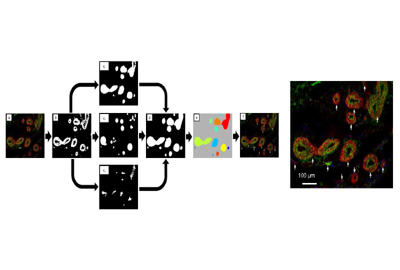
Tema: NET-IBA, sviluppo di algoritmi e metodi automatici per l’analisi strutturale e morfologica di tessuti nativi e scaffolds. L’istopatologia ed i metodi di analisi quantitativa di struttura su base immagine sono al momento scarsamente sviluppati in modo integrato. Gran parte delle valutazioni in istologia avvengono ancora sulla base di stime qualitative o semi-qualitative. Analogamente, i metodi di analisi strutturale sulla base di immagine, sviluppati in contesti di scienza dei materiali, trascurano svariate metriche quantitative per micro e meso architettura. Questa linea di ricerca si posiziona all’interfaccia tra queste due discipline nel tentativo di fornire nuovi strumenti di indagine sia in ambito clinico che in ambito scienza dei materiali.
Impact:
The software tools we developed and that we are tryaing to advance have the potential to impact on two main categories of problems:
– quantitative histology, potential applications include: biomaterial-host interactions, evaluation of drugs effects on tissue, inflammatory response evaluation, oncology, tissue elaboration in vitro and in vivo, big data;
– morphological analysis of micro and nano-structured materials, potential applications within the context of chemical, process engineering or material science, include: process control, process characterization, structure-function characterization.
Pipeline
-
CLINICAL
NEED -
DISEASES
ANALYSIS - DISCOVERY
-
PRECLINICAL
VALIDATION -
PRECLINICAL
DEVELOPMENT -
CLINICAL
STUDIES
Principal Investigator
Contact
Team di progetto:
Therapeutic area:
Products:
ATMP – Medical devices & tissue engineering
Collaborations:
University of Pittsburgh, PA
Università degli Studi di Palermo, Italia
Scarica il pdf del progetto